-
By:
- jayson
- No comment
image guided srt for skin cancer
Image-Guided Stereotactic Radiotherapy (SRT) for Skin Cancer: A Comprehensive Overview
Image-guided SRT represents a non-invasive advancement, precisely targeting skin cancers with focused radiation beams, offering a compelling alternative to conventional methods.
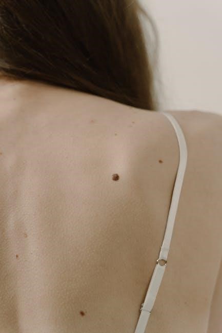
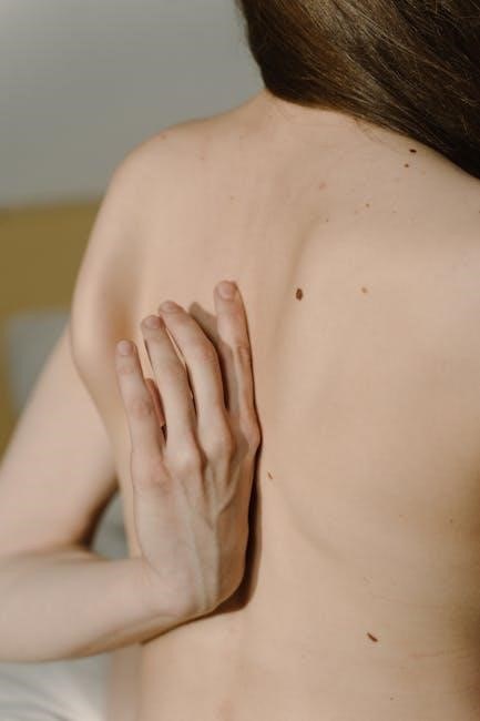
Skin cancer is the most common malignancy, impacting millions globally, with basal cell carcinoma, squamous cell carcinoma, and melanoma being the primary types. Traditional treatments – surgical excision, Mohs surgery, and conventional radiation therapy – have long been the standards of care. However, advancements continually refine treatment precision and minimize side effects.
Image-guided Stereotactic Radiotherapy (SRT) emerges as a sophisticated, non-invasive option, particularly beneficial for patients unsuitable for surgery or seeking alternatives. SRT delivers highly focused radiation doses to the tumor while sparing surrounding healthy tissues. This technique leverages advanced imaging to pinpoint the cancer’s location with sub-millimeter accuracy, ensuring effective treatment and improved cosmetic outcomes. Exploring these modern approaches is crucial for optimizing patient care and quality of life.
Understanding Stereotactic Radiotherapy (SRT)
Stereotactic Radiotherapy (SRT) is a highly precise form of radiation therapy that delivers a large dose of radiation to a small, well-defined target volume. Unlike conventional radiation, SRT utilizes sophisticated imaging techniques to accurately locate and target the tumor, minimizing exposure to surrounding healthy tissues. This is achieved through meticulous treatment planning and real-time image guidance.
The core principle of SRT revolves around delivering a hypofractionated dose – meaning fewer, larger radiation fractions – over a short period. This approach is biologically equivalent to traditional fractionation but offers convenience and potentially reduces treatment-related toxicity. SRT’s precision makes it particularly suitable for skin cancers in cosmetically sensitive areas or near critical structures, offering a compelling alternative to surgical intervention.
How SRT Differs from Traditional Radiation Therapy
Traditional radiation therapy typically employs multiple, smaller radiation fractions delivered over several weeks, aiming to gradually control tumor growth. In contrast, SRT utilizes fewer, larger doses delivered with pinpoint accuracy over a condensed timeframe – often just one to five sessions.
A key distinction lies in the technology. SRT leverages advanced imaging – like CT, MRI, or PET/CT – for precise tumor localization and treatment planning. Traditional methods often rely on less detailed imaging. Furthermore, SRT incorporates real-time image guidance during treatment to account for patient movement and anatomical changes, ensuring the radiation beam remains focused on the target. This heightened precision minimizes damage to healthy tissue and improves treatment efficacy.
The Principles of Precise Radiation Delivery
Precise radiation delivery in SRT hinges on several core principles. First, highly focused radiation beams, often shaped using techniques like multi-leaf collimation (MLC), conform closely to the tumor’s shape, sparing surrounding healthy tissues. Second, sophisticated treatment planning systems calculate the optimal beam angles and intensities to maximize the dose to the tumor while minimizing exposure to critical structures.
Third, immobilization devices – custom masks or molds – ensure the patient remains in a consistent position throughout treatment. Finally, real-time image guidance, typically using Cone-Beam CT (CBCT), verifies the tumor’s location before and during each fraction, allowing for adjustments to maintain accuracy. These combined elements ensure a highly targeted and effective radiation dose.
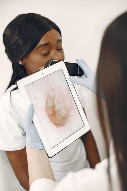
The Role of Imaging in SRT for Skin Cancer
Imaging is absolutely central to the success of SRT for skin cancer, extending far beyond initial diagnosis. It’s crucial for accurate tumor delineation, defining the precise boundaries of the cancerous tissue and surrounding critical structures. Detailed imaging guides treatment planning, allowing clinicians to create a personalized radiation plan that maximizes tumor control while minimizing damage to healthy skin and underlying tissues.
Furthermore, imaging plays a vital role in real-time treatment verification. Before each fraction, imaging confirms the tumor’s position hasn’t shifted due to breathing or patient movement. This ensures the radiation is delivered to the intended target with sub-millimeter accuracy, a cornerstone of SRT’s effectiveness.
Importance of Accurate Tumor Localization
Precise tumor localization is paramount in image-guided SRT for skin cancer, directly impacting treatment efficacy and minimizing side effects. Skin cancers, particularly those on mobile areas like the face, can shift with even minor movements. Accurate localization ensures the radiation beam consistently targets the tumor volume, maximizing the dose delivered to cancerous cells while sparing surrounding healthy tissue.
Even slight inaccuracies can lead to under-dosing the tumor, potentially causing recurrence, or over-dosing healthy tissues, increasing the risk of complications. Advanced imaging techniques and real-time verification systems are therefore essential to account for physiological movements and anatomical variations, guaranteeing treatment precision.
Imaging Modalities Used: CT, MRI, and PET/CT
Several imaging modalities play crucial roles in image-guided SRT for skin cancer. Computed Tomography (CT) provides high-resolution anatomical detail, essential for treatment planning and accurate dose calculation. Magnetic Resonance Imaging (MRI) offers superior soft tissue contrast, particularly valuable for assessing tumor extent and involvement of nearby structures.
Positron Emission Tomography/Computed Tomography (PET/CT) combines functional and anatomical imaging, aiding in detecting microscopic disease and differentiating between scar tissue and residual cancer. The choice of imaging modality depends on the specific cancer type, location, and clinical scenario. Often, a combination of these techniques is utilized to achieve the most comprehensive assessment and guide precise radiation delivery;
Patient Selection Criteria for Image-Guided SRT

Careful patient selection is paramount for successful image-guided SRT. Ideal candidates generally include those with histologically confirmed skin cancers – basal cell carcinoma, squamous cell carcinoma, and select melanomas – that are not amenable to surgical resection due to location, size, or patient comorbidities.
Factors considered include tumor depth, proximity to critical structures, prior treatment history, and overall health status. Patients with significant medical conditions or those unable to remain still during treatment may not be suitable. A thorough evaluation, including a comprehensive medical history and physical examination, is essential to determine appropriateness and maximize treatment outcomes.
Suitable Skin Cancer Types (Basal Cell, Squamous Cell, Melanoma)
Image-guided SRT demonstrates efficacy across a spectrum of skin cancer types. Basal cell carcinomas (BCCs), particularly those in cosmetically sensitive areas or recurrent lesions, respond exceptionally well; Squamous cell carcinomas (SCCs), especially those with higher risk features, are also frequently treated with SRT.
Melanoma, specifically thin melanomas in situ or with minimal depth of invasion, can be considered for SRT, though careful staging and follow-up are crucial. The suitability depends on histological subtype, tumor location, and patient-specific factors. SRT offers a non-surgical option, preserving functionality and cosmetic outcomes.
Tumor Characteristics & Anatomical Considerations
Successful SRT planning hinges on meticulous assessment of tumor characteristics. Tumor size and depth are critical; SRT is generally most effective for smaller, well-defined lesions. Proximity to critical structures – eyes, nose, cartilage – significantly influences dose planning and treatment feasibility.
Anatomical location also plays a role. Areas prone to movement, like the nasolabial folds, require robust immobilization. Histological features, such as perineural invasion, may impact treatment decisions. Careful consideration of these factors ensures optimal targeting and minimizes potential side effects, maximizing treatment efficacy.
The SRT Treatment Planning Process

The SRT treatment journey begins with detailed planning, utilizing high-resolution imaging – typically CT scans – to create a three-dimensional model of the treatment area. A radiation oncologist meticulously contours the tumor and surrounding critical structures. Dose calculations are then performed to determine the optimal radiation dose distribution, maximizing tumor control while sparing healthy tissue.

Beam angles and shapes are carefully selected to conform to the tumor’s geometry. The plan is rigorously reviewed and approved before treatment commences. Sophisticated software aids in visualizing the planned dose distribution, ensuring accuracy and safety. This iterative process is crucial for personalized, effective SRT.
Immobilization and Masking Techniques
Accurate and consistent patient positioning is paramount in SRT. Immobilization devices, often custom-fitted masks, are crucial for minimizing movement during treatment. These masks, typically made of thermoplastic material, conform precisely to the patient’s facial contours, ensuring reproducible setup. The mask is secured to the treatment table, preventing shifts that could compromise accuracy.
For areas beyond the head and neck, specialized body frames or vacuum-lock cushions may be employed. Regular verification with imaging confirms proper alignment before each fraction. Patient comfort is also considered during mask fabrication and treatment sessions. Effective immobilization is fundamental to delivering the prescribed dose precisely to the target.
Dose Calculation and Beam Shaping
Precise dose calculation is central to SRT’s efficacy. Treatment planning systems utilize detailed imaging data to map the tumor’s location and surrounding tissues. Sophisticated algorithms then determine the optimal radiation dose distribution, maximizing tumor control while sparing healthy skin and underlying structures. Beam shaping techniques, like multi-leaf collimation (MLC), conform the radiation beam precisely to the tumor’s contours.
Intensity-modulated radiation therapy (IMRT) further refines beam delivery, allowing for non-uniform dose distributions. Dose constraints are carefully established to limit radiation exposure to critical organs. Rigorous quality assurance procedures verify the accuracy of the treatment plan before implementation, ensuring safe and effective therapy.
Real-Time Image Guidance During Treatment
Real-time image guidance is a cornerstone of SRT’s accuracy. Before each radiation fraction, imaging is performed to verify the patient’s position and any potential anatomical changes since the initial treatment plan. This ensures the radiation beam consistently targets the intended tumor volume. Image guidance systems detect even subtle movements, allowing for adjustments before radiation delivery.
This dynamic adaptation minimizes the risk of targeting healthy tissue and maximizes the dose delivered to the cancer. Automated patient positioning systems, coupled with image verification, enhance treatment precision. Continuous monitoring throughout the treatment session further refines accuracy, providing a robust safety net.
Utilizing Cone-Beam CT (CBCT) for Verification
Cone-Beam Computed Tomography (CBCT) is a crucial imaging technique employed during SRT treatment verification. Unlike traditional CT scans, CBCT delivers a lower radiation dose while providing high-resolution, 3D images of the treatment area. This allows for precise assessment of the patient’s anatomy immediately before each radiation fraction.
CBCT imaging confirms that the tumor remains within the planned target volume and identifies any shifts caused by breathing, weight loss, or patient movement. Any discrepancies detected are corrected by adjusting the patient’s position or modifying the treatment plan. This iterative process ensures the radiation is delivered with pinpoint accuracy, maximizing efficacy and minimizing side effects.
Addressing Patient Movement and Anatomical Changes
Patient movement during treatment and subtle anatomical changes between fractions pose significant challenges in SRT. Respiratory motion, even minor shifts in position, can compromise treatment accuracy. Image guidance, particularly with CBCT, plays a vital role in mitigating these issues.
Real-time monitoring and image-based verification allow for adjustments to the treatment setup before each beam delivery. Automated couch corrections are frequently employed to align the patient with the original treatment plan. Furthermore, adaptive radiotherapy techniques can modify the beam shape or dose distribution to account for anatomical changes. Proactive management of these factors is essential for optimal treatment outcomes and minimizing damage to surrounding healthy tissues.
Benefits of Image-Guided SRT for Skin Cancer
Image-guided SRT offers several distinct advantages over traditional radiation and even surgical excision for select skin cancers. Reduced treatment time is a key benefit, often requiring fewer sessions – typically one to five – compared to weeks of conventional radiotherapy. This convenience improves patient compliance and quality of life.
Precise targeting minimizes radiation exposure to surrounding healthy tissues, reducing the risk of long-term side effects. Non-invasive nature avoids surgical scars and associated complications. Furthermore, SRT is particularly beneficial for patients with tumors in cosmetically sensitive areas or those who are not surgical candidates due to age or comorbidities. Improved cosmetic outcomes and a favorable toxicity profile contribute to its growing popularity.
Reduced Treatment Time & Fewer Sessions
A significant advantage of image-guided SRT lies in its ability to deliver a high dose of radiation precisely to the tumor in a limited number of fractions. Unlike conventional radiotherapy, which typically spans several weeks with daily treatments, SRT often completes treatment in just one to five sessions.
This condensed schedule dramatically reduces the overall treatment burden for patients, minimizing disruption to their daily lives. Shorter treatment courses also translate to lower healthcare costs and decreased travel requirements. The ability to achieve equivalent tumor control with fewer sessions enhances patient convenience and compliance, ultimately improving treatment outcomes and satisfaction.
Minimizing Damage to Surrounding Healthy Tissue
Image-guided SRT excels in its precision, a key factor in sparing healthy tissue surrounding the skin cancer. The sophisticated imaging techniques used – like CBCT – allow for real-time adjustments, ensuring the radiation beam conforms precisely to the tumor’s shape and location.
This targeted approach significantly reduces the dose delivered to adjacent normal tissues, minimizing potential side effects. By concentrating the radiation on the cancerous cells, SRT preserves the function and appearance of the treated area, leading to improved cosmetic outcomes and a better quality of life for patients. Reduced toxicity is a cornerstone of this advanced treatment modality.

Potential Side Effects and Management
While generally well-tolerated, image-guided SRT for skin cancer can cause some side effects. Common reactions include skin redness, dryness, and mild discomfort at the treatment site, resembling a sunburn. These are typically temporary and manageable with moisturizers and gentle wound care.
Less frequent side effects may involve skin breakdown or, rarely, deeper tissue effects. Proactive management, including diligent skin monitoring and following post-treatment instructions, is crucial. Patients are educated on recognizing and reporting any concerning symptoms to their care team, ensuring prompt intervention and optimal healing. Open communication is key to minimizing discomfort and maximizing treatment success.
Common Skin Reactions & Wound Care
Following SRT, patients commonly experience skin reactions similar to a mild to moderate sunburn – redness, warmth, and potential dryness. These are expected responses and usually subside within a few weeks. Gentle wound care is paramount; avoid harsh soaps, scrubbing, or direct sun exposure.
Moisturizing with fragrance-free, hypoallergenic creams is highly recommended to maintain skin integrity. Non-adhesive dressings may be used to protect the treated area, especially if blistering occurs. Patients should avoid picking at any scabs or blisters to prevent infection and promote optimal healing. Promptly report any signs of infection, such as increased pain, swelling, or pus, to their physician.
Long-Term Effects & Monitoring
While SRT boasts a high cure rate, long-term monitoring is crucial. Late effects are uncommon but can include skin changes like hyperpigmentation or telangiectasias (small, visible blood vessels). Rarely, radiation-induced fibrosis may occur, causing tissue hardening.
Regular follow-up appointments, typically every 3-6 months for the first two years, then annually, are essential. These visits involve clinical skin examinations to assess for recurrence or new primary skin cancers. Patients should also perform self-skin exams regularly and report any new or changing lesions to their dermatologist. Ongoing surveillance ensures early detection and management of any potential long-term complications.
SRT vs. Surgical Excision: A Comparative Analysis
Surgical excision remains the gold standard for many skin cancers, offering immediate tumor removal and pathological confirmation. However, SRT presents a compelling non-invasive alternative, particularly for patients unsuitable for surgery or desiring cosmetic preservation.
SRT generally results in less scarring and preserves surrounding tissue architecture better than excision. Recurrence rates are comparable between the two modalities when appropriately indicated. Surgical excision requires local anesthesia and potential wound care, while SRT is painless and requires no incisions. The choice depends on tumor size, location, histology, patient preference, and overall health.
The Future of Image-Guided SRT in Skin Cancer Treatment
Ongoing research focuses on refining SRT techniques, including adaptive planning based on real-time tumor response and incorporating artificial intelligence for automated contouring and dose optimization. Hypofractionation protocols are being explored to further reduce treatment time without compromising efficacy.
Combining SRT with immunotherapy is a promising avenue, potentially enhancing anti-tumor immune responses. Development of novel imaging agents will improve tumor visualization and treatment monitoring. Ultimately, the future envisions personalized SRT regimens tailored to individual patient and tumor characteristics, maximizing outcomes and minimizing side effects, establishing SRT as a cornerstone of skin cancer care.