-
By:
- jayson
- No comment
essentials of human anatomy and physiology laboratory manual
This manual provides hands-on learning experiences, combining essential concepts of anatomy and physiology with practical skills, enabling students to explore the human body’s structure and functions effectively.
Overview of the Laboratory Manual
This laboratory manual is designed to complement anatomy and physiology coursework, offering a structured approach to hands-on learning. It includes detailed exercises, clear instructions, and visual aids to guide students through experiments. Each lab activity is tailored to reinforce key concepts, from skeletal and muscular systems to nervous system functions. The manual emphasizes safety protocols and proper use of equipment, ensuring a secure learning environment. It also integrates with accompanying textbooks, providing a comprehensive resource for understanding human anatomy and physiology. Through practical exercises, students gain the ability to apply theoretical knowledge, fostering a deeper understanding of biological processes. This manual serves as an essential tool for engaging with complex topics in a structured and accessible manner.
Importance of Hands-On Learning in Anatomy and Physiology
Hands-on learning is crucial for mastering anatomy and physiology, as it allows students to engage directly with biological structures and processes. Practical exercises enable a deeper understanding of complex concepts, enhancing retention and critical thinking. By participating in lab activities, students can visualize and interact with anatomical structures, making abstract ideas more tangible. This approach also fosters problem-solving skills and collaboration, simulating real-world scenarios in healthcare and research. The laboratory environment provides a safe space for experimentation and exploration, where students can apply theoretical knowledge to practical tasks. Hands-on learning not only strengthens comprehension but also builds confidence in manipulating tools and techniques essential for future careers in medicine, nursing, and allied health fields. It bridges the gap between textbook learning and real-world applications, making it an indispensable component of anatomy and physiology education.
Essential Laboratory Skills and Safety Protocols
Mastery of microscopy, dissection, and measurement techniques is vital. Adhering to safety protocols ensures a secure environment, preventing accidents and fostering a focus on precise, efficient lab practices.
Setting Up the Laboratory Environment
Setting up the laboratory environment involves organizing workstations, ensuring cleanliness, and arranging essential tools. Begin by cleaning and disinfecting all surfaces and equipment. Position microscopes, dissection tools, and gloves within easy reach. Label specimen containers clearly and assign storage areas for supplies. Ensure proper ventilation and access to safety equipment like fire extinguishers and first aid kits. Designate zones for waste disposal, separating biohazardous materials from general trash. Familiarize yourself with emergency exits and procedures. Finally, conduct a walkthrough to verify all systems are ready for safe and efficient experimentation.
- Workspace preparation ensures clarity and focus.
- Essential tools like microscopes and gloves must be accessible.
- Clear labeling and organized storage prevent confusion.
Using Microscopy and Dissection Tools
Mastering microscopy and dissection tools is fundamental in anatomy and physiology labs. Begin by preparing slides for microscopy, ensuring specimens are properly stained and mounted. Focus the microscope by adjusting the condenser and objective lenses, starting with low magnification before switching to higher powers. For dissection, select appropriate tools like scalpels, forceps, and probes, and practice precise cuts to avoid damaging tissues. Always handle sharp instruments with care and store them safely when not in use. Regularly clean and maintain equipment to ensure optimal performance. Proper techniques enhance observation and exploration of anatomical structures, fostering a deeper understanding of physiological processes.
- Microscopy reveals cellular details essential for analysis.
- Dissection tools require skill and precision for effective use.
Human Anatomy Laboratory Exercises
Engage in hands-on exploration of the human body through structured exercises, examining skeletal, muscular, and organ systems to gain practical insights into anatomical structures and their functions.
Exploring the Skeletal System: Bones and Joints
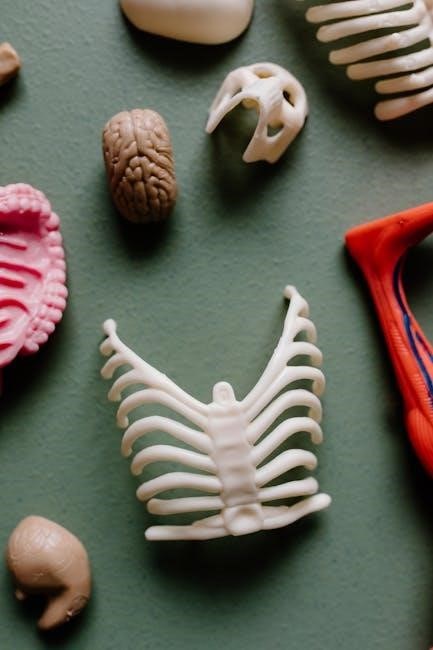
The skeletal system forms the structural framework of the human body, comprising 206 bones and their associated joints. This section of the manual focuses on identifying and understanding the classification of bones based on their shapes—long, short, flat, irregular, and sesamoid. Through hands-on activities, students examine the unique features of each bone type, such as the shaft (diaphysis) and ends (epiphyses) of long bones, and the spongy tissue within flat and irregular bones. Joints, or articulations, are also explored, with an emphasis on their types (synovial, cartilaginous, and fibrous) and functions in facilitating movement. Practical exercises include labeling bone models, observing joint structures, and simulating movements to understand the relationship between bones and joints. These activities provide a foundational understanding of how the skeletal system supports mobility and stability in the body, essential for further studies in anatomy and physiology.
Identifying Muscular Structures and Their Functions
This section focuses on the detailed study of the muscular system, emphasizing the identification and functional analysis of major muscle groups. Students learn to distinguish between skeletal, smooth, and cardiac muscles, understanding their roles in voluntary and involuntary movements. Hands-on activities include dissecting specimens to observe muscle attachments and fiber arrangements. Practical exercises involve palpating muscles on the human body to correlate anatomical structures with physical sensations. The manual also covers the classification of muscles based on their actions, such as flexors, extensors, and rotators. By exploring muscle physiology, students gain insights into how muscles interact with bones and joints to produce movement. These exercises enhance understanding of muscle function and prepare students for advanced studies in kinesiology and physical therapy, bridging anatomy with real-world applications in health and medicine.
Physiology Laboratory Experiments
Physiology laboratory experiments involve measuring vital signs, such as heart rate and blood pressure, and conducting reflex tests to understand nervous system responses and overall physiological processes.
Measuring Vital Signs: Heart Rate, Blood Pressure, and Respiration
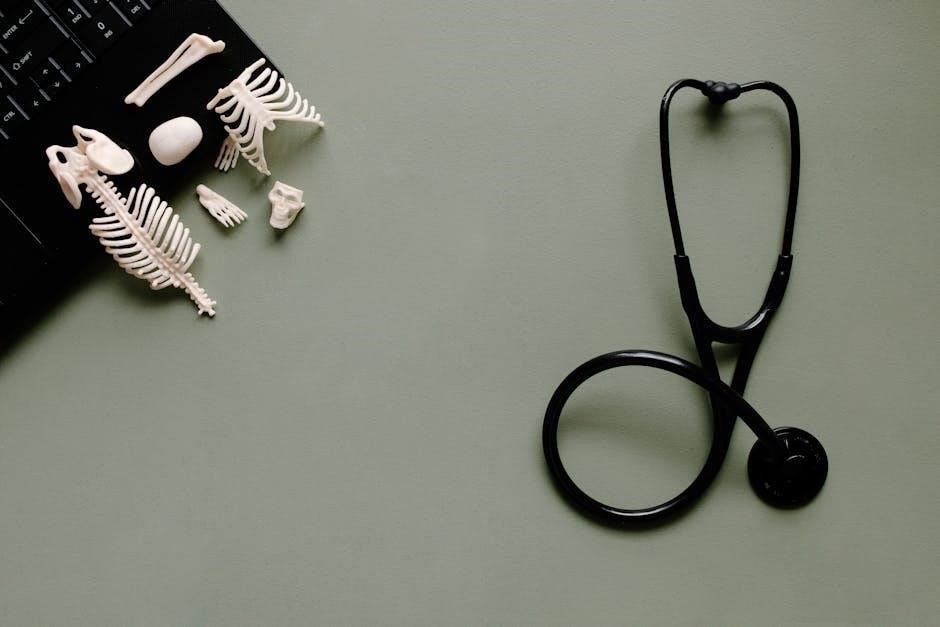
Measuring vital signs is a fundamental skill in anatomy and physiology labs. Heart rate, typically ranging from 60 to 100 beats per minute, indicates cardiac efficiency. Blood pressure, measured in millimeters of mercury, reflects cardiovascular health, with normal readings around 120/80 mmHg. Respiration rate, usually 12-20 breaths per minute, assesses pulmonary function. These measurements provide baseline data for understanding physiological responses to various conditions. Students learn to use stethoscopes and sphygmomanometers to accurately record these metrics, essential for diagnosing health statuses and monitoring changes during experiments. This hands-on practice reinforces the importance of precise measurement in healthcare and research, preparing students for real-world applications in medical fields.
Understanding Nervous System Responses Through Reflex Testing
Reflex testing is a critical component of physiology labs, allowing students to explore the nervous system’s role in controlling involuntary actions. By eliciting reflexes, such as the knee-jerk response, students observe how sensory input triggers motor output through neural pathways. This hands-on approach demonstrates the integration of the nervous system in maintaining homeostasis and responding to stimuli. Labs often involve testing stretch reflexes and withdrawal reflexes, using tools like reflex hammers to initiate responses. Observing these reactions provides insights into the nervous system’s functionality, including the speed of neural signaling and the body’s adaptive mechanisms. Such experiments are vital for understanding how the nervous system coordinates and regulates bodily functions, offering practical applications in clinical diagnostics and therapeutic interventions.
This comprehensive laboratory manual effectively integrates anatomy and physiology through hands-on experiments, fostering a deep understanding of human biology and its practical applications in real-world scenarios.
Key Takeaways from the Laboratory Manual
The laboratory manual emphasizes the integration of anatomy and physiology through hands-on experiments, enhancing understanding of human body systems. Students gain practical skills in microscopy, dissection, and data collection, fostering critical thinking. The manual’s structured format, with clear objectives and visual aids, ensures comprehensive learning. Key concepts include skeletal and muscular systems, nervous system responses, and vital sign measurement. Real-world applications of lab exercises prepare students for clinical scenarios, bridging theory and practice. The manual’s focus on essential experiments and safety protocols equips learners with foundational skills for future careers in healthcare and science.
Applying Laboratory Knowledge to Real-World Scenarios
Laboratory experiences in anatomy and physiology directly translate to real-world healthcare applications, enabling students to understand clinical practices. Measuring vital signs, such as heart rate and blood pressure, prepares learners for patient assessments in medical settings. Understanding skeletal and muscular systems aids in diagnosing injuries or conditions like fractures or muscle strains. Reflex testing provides insights into nervous system function, crucial for neurological evaluations. Additionally, skills in microscopy and tissue identification are vital in pathology and research. The manual’s focus on enteral feeding and tube management equips students for roles in nutrition and patient care. By bridging theoretical knowledge with practical skills, the laboratory manual prepares future professionals to address diverse medical scenarios effectively.